Bursitis Of The Foot Operations
Overview
Plantar calcaneal bursitis is a medical condition in which there is inflammation of the plantar calcaneal bursa, a spongy fluid filled sac that cushions the fascia of the heel and the calcaneus (heel bone). It is characterized by swelling and tenderness of the central plantar heel area. It is sometimes called 'Policeman's heel'. It sometimes was, and should not be, confused with plantar fasciitis, which is inflammation of the plantar fascia and can affect any part of the foot.
Causes
The swelling is the result of the blockage of blood, tissue fluids and circulation in the heel because their normal movement has been disrupted by the force of the injury. Just like cars back up behind a traffic jam, causing congestion, exhaust and overheating, blood and fluids back up behind the injured heel, causing pain, inflammation, lumps and swelling.
Symptoms
Some of the symptoms of bursitis in the heel, or retrocalcaneal bursitis, are as described below. Severe pain in the heel area of the foot, sometimes radiating to the ankle, associated with physical activities like walking, jogging and even on physical contact to the area. The physical signs of heel bursitis, which are noticeable in the heel area, are reddish discoloration of the skin that is warm to touch.
Diagnosis
Diagnosis of heel bursitis can be made by your health practitioner and is based on the following. Assessing the location of the pain by palpating the back of the heel. Assessment of any inflammation at the back of the heel. Assessment of biomechanics and foot function. Ultrasound or MRI can reveal inflammation of the retro calcaneal bursa.
Non Surgical Treatment
Many times, Achilles tendon bursitis can be treated with home care. However, if self-care remedies do not work, your doctor may need to administer additional treatments. Home care. The most important thing you can do to help your healing is to refrain from activities that could further aggravate the bursitis. Typical home-care treatments for Achilles tendon bursitis include Anti-inflammatory medication. Take over-the-counter pain medication, like aspirin or ibuprofen, to reduce pain and swelling. Heel wedges. An over-the-counter or custom heel wedge can be placed in your shoe to minimize stress in the Achilles tendon. Ice. Apply ice or other cold therapy to your sore heel several times a day. Rest. Limit your activity on the injured leg, avoid putting pressure on it whenever possible. Also, elevate your leg during non-use to help reduce the swelling. The above remedies may be used on their own or in combination with others on the list. Physician-administered treatments. If the above self-care remedies are not effective, you should visit your doctor for additional treatment. Possible Achilles tendon bursitis treatments your doctor may administer include immobilization. If the bursitis is combined with Achilles tendonitis, your doctor may place a temporary cast on the ankle to prevent movement and allow it to heal. Physical therapy. Exercises may be used to improve the ankle's strength and flexibility. Steroids. Injection of steroids into the retrocalcaneal bursa (not the Achilles tendon) may be necessary. Surgery. In very rare circumstances, surgery may be needed to remove the retrocalcaneal bursa, however, this is typically a last resort.
Surgical Treatment
Surgery to remove the damaged bursa may be performed in extreme cases. If the bursitis is caused by an infection, then additional treatment is needed. Septic bursitis is caused by the presence of a pus-forming organism, usually staphylococcus aureus. This is confirmed by examining a sample of the fluid in the bursa and requires treatment with antibiotics taken by mouth, injected into a muscle or into a vein (intravenously). The bursa will also need to be drained by needle two or three times over the first week of treatment. When a patient has such a serious infection, there may be underlying causes. There could be undiscovered diabetes, or an inefficient immune system caused by human immunodeficiency virus infection (HIV).
Prevention
After taking a history and performing a physical examination, your physician may order x-rays to rule out other disorders. Your doctor may administer injections of corticosteroids and a local anesthetic to reduce swelling and ease pain. Also, to reduce swelling, your physician may draw excess fluid from the bursa with a syringe and then tightly wrap and compress the joint with an elastic bandage. In severe, persistent cases surgery to remove the bursa may be necessary. For infectious bursitis, antibiotics will be prescribed.
HammerToes
 Overview
Uneven muscle tension results in the distortion of one or several of the small toes. (Hammer toe) Pressure points develop at the raised middle joint as well as at the tip of the toe and underneath the metatarsal head. In the beginning, when the misalignment can still be corrected, it often suffices to lengthen the tendon and to cut a notch into the capsule. In a hammertoes contracted misalignment, part of the middle joint is removed to form a replacement joint. Modern surgical techniques preserve the metatarsophalangeal joint (Weil or Helal osteotomies).
Causes
Most hammertoes are caused by wearing ill-fitting, tight or high-heeled shoes over a long period of time. Shoes that don?t fit well can crowd the toes, putting pressure on the middle toes and causing them to curl downward. Other causes include genes. Some people are born with hammertoe, bunions. These knobby bumps sometimes develop at the side of the big toe. This can make the big toe bend toward the other toes. The big toe can then overlap and crowd the smaller toes. Arthritis in a toe joint can lead to hammertoe.
 Symptoms
Hammer toe is often distinguished by a toe stuck in an upside-down ?V? position, and common symptoms include corns on the top of your toe joint. Pain at the top of a bent toe when you put on your shoes. Pain when moving a toe joint. Pain on the ball of your foot under the bent toe. Corns developing on the top of the toe joint. It is advisable to seek medical advice if your feet hurt on a regular basis. It is imperative to act fast and seek the care of a podiatrist or foot surgeon. By acting quickly, you can prevent your problem from getting worse.
Diagnosis
Your doctor is very likely to be able to diagnose your hammertoe simply by examining your foot. Even before that, he or she will probably ask about your family and personal medical history and evaluate your gait as you walk and the types of shoes you wear. You'll be asked about your symptoms, when they started and when they occur. You may also be asked to flex your toe so that your doctor can get an idea of your range of motion. He or she may order x-rays in order to better define your deformity.
Non Surgical Treatment
For hammertoes that are still flexible, a podiatrist might recommend padding or taping the toes to relieve pain and orthotic inserts for shoes to minimize pressure and keep the toe properly aligned. Anti-inflammatory drugs or corticosteroid injections can relieve pain and inflammation. For more advanced cases of hammertoe, a podiatrist might recommend a surgical procedure to cut the tendon, allowing the toe to straighten. For hammertoes that have become rigid, a more complicated surgery might be needed, during which the podiatrist removes part of the bone at the deformed joint to allow it to straighten.
Surgical Treatment
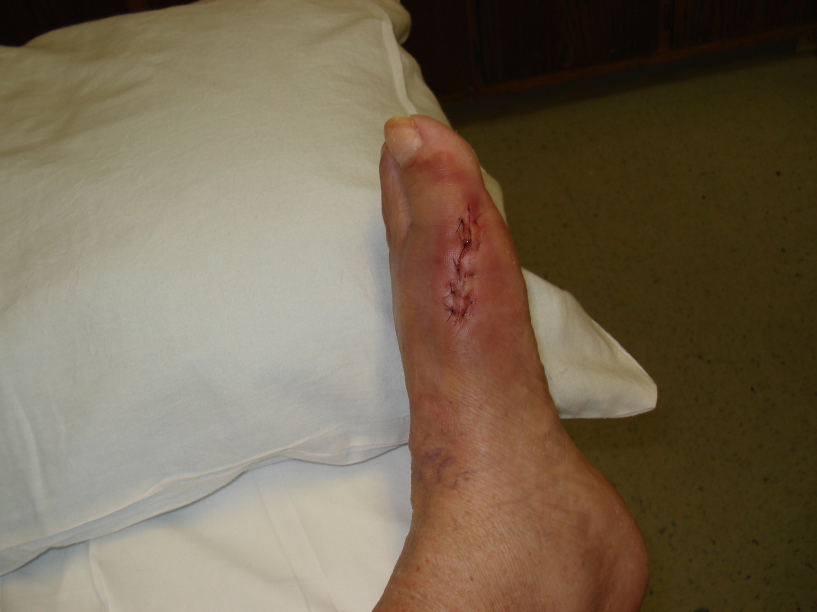
In more advanced cases of hammer toe, or when the accompanying pain cannot be relieved by conservative treatment, surgery may be required. Different types of surgical procedures are performed to correct hammer toe, depending on the location and extent of the problem. Surgical treatment is generally effective for both flexible and fixed (rigid) forms of hammer toe. Recurrence following surgery may develop in persons with flexible hammer toe, particularly if they resume wearing poorly-fitted shoes after the deformity is corrected.
Hammer Toe Pain After Surgery
 Overview
Hammer toe is defined as a deformity in the toe where part of the toe is permanently bent downward resembling a hammer. Two related conditions are mallet toe and claw toe which effect different toe joints in slightly different ways. The key difference is that hammertoes tends to effect the middle joint in the toe (note: not the middle toe, the middle toe joint). The disease is usually associated with the second largest toe but can effect the third or fourth toe as well. Mallet toe effects the uppermost toe joint whereas claw toe is caused by the tow being held in a cramped ?claw-like? position.
Causes
More often than not, wearing shoes that do Hammer toe not fit a person well for too long may actually cause hammer toes. Wearing shoes that are too narrow or too tight for the person for extended periods of time may eventually take a toll on the person's feet. The same is true for women who like wearing high-heeled shoes with narrow toe boxes.
 Symptoms
Pain on the bottom of your foot, especially under the ball of your foot, is one of the most common symptoms associated with hammertoes. Other common signs and symptoms of hammertoes include pain at the top of your bent toe from footwear pressure. Corns on the top of your bent toe. Redness and swelling in your affected area. Decreased joint range of motion in your affected toe joints.
Diagnosis
Most health care professionals can diagnose hammertoe simply by examining your toes and feet. X-rays of the feet are not needed to diagnose hammertoe, but they may be useful to look for signs of some types of arthritis (such as rheumatoid arthritis) or other disorders that can cause hammertoe. If the deformed toe is very painful, your doctor may recommend that you have a fluid sample withdrawn from the joint with a needle so the fluid can be checked for signs of infection or gout (arthritis from crystal deposits).
Non Surgical Treatment
A person with hammer toes will be asked to practice some exercises for their toes to regain average structure and movement. The exercises usually involve stretching and strengthening their toes. The person may attempt to pick things up off the floor using only their toes. They may also stretch their toes on a regular basis by hand to ease them into straightening out. Another example of a physical exercise specifically for a person's toes involves crumpling a towel with the toes. The towel can lie underneath the person's feet and the person can use their toes to scrunch up the towel as they perform simple tasks such as reading a book or watching television.
Surgical Treatment
Surgically correcting a hammertoe is very technical and difficult, and requires a surgeon with superior capabilities and experience. The operation can be done at our office or the hospital with local anesthetic. After making a small incision, the deformity is reduced and the tendons are realigned at the joint. You will be able to go home the same day with a special shoe! If you are sick and tired of not fitting your shoes, you can no longer get relief from pads, orthopedic shoes or pedicures, and have corns that are ugly, sensitive and painful, then you certainly may be a good surgical candidate. In order to have this surgery, you can not have poor circulation and and must have a clean bill of health.
 Prevention
Certain exercises such as moving and stretching your toe gently with your hands and picking up small or soft objects such as marbles or towels can keep your toe joints flexible, simple exercises can stretch and strengthen your muscles. Limit high-heel use, well-designed flat shoes will be more comfortable than high heels. Don't wear shoes that are too short or too narrow, or too shallow, this is especially important for children going through periods of rapid growth, the toe area should be high enough so that it doesn't rub against the top of your toes.
Over-Pronation Of The Feet What Are The Causes
Overview
Overpronation is the most common abnormality found in the foot, and for this reason, is the most studied. The term that most people attribute to overpronation is ?flat feet.? Pronation is the rolling in of the foot and the collapse of the arch. Every person pronates to some extent and this is a necessary moment in the normal walking cycle as it allows the forefoot to make complete contact with the ground. A foot that overpronates acts like a loose bag of bones during the walking cycle. This makes this type of foot very flexible but inefficient. The foot has to work much harder to propel the body, fatiguing easily and placing mechanical stresses on the lower body. We like to use the analogy of digging a hole in the dirt. Overpronating feet are like using a broom to dig the hole. It won?t break down quickly, but you will be digging for a very long time, or until eventually wear and tear will take effect. Wouldn?t you rather have a shovel to work with. This is in essence what an orthotic can do for your feet. This is why orthotics have become an evidence based treatment for so many foot ailments, as they can effectively manage overpronation.Overpronation is when a person pronates too much and for too long. This places excess stress on the tendons and ligaments in the foot and ankle.
Causes
It is important to identify the cause of overpronation in order to determine the best treatment methods to adopt. Not all treatments and preventative measures will work equally well for everyone, and there may be a little trial and error involved to get the best treatment. A trip to a podiatrist or a sports therapist will help you to establish the cause of overpronation, and they will be able to tell you the best treatments based on your specific degree of overpronation and the cause. Overpronation has many causes, with the most common reasons for excessive pronation listed, low arches, flexible flat feet, fallen arches, gait abnormalities, abnormal bone structure, abnormal musculature, bunions, corns and calluses.
Symptoms
Because pronation is a twisting of the foot, all of the muscles and tendons which run from the leg and ankle into the foot will be twisted. In over-pronation, resulting laxity of the soft tissue structures of the foot and loosened joints cause the bones of the feet shift. When this occurs, the muscles which attach to these bones must also shift, or twist, in order to attach to these bones. The strongest and most important muscles that attach to our foot bones come from our lower leg. So, as these muscles course down the leg and across the ankle, they must twist to maintain their proper attachments in the foot. Injuries due to poor biomechanics and twisting of these muscles due to over-pronation include: shin splints, Achilles Tendonitis, generalized tendonitis, fatigue, muscle aches and pains, cramps, ankle sprains, and loss of muscular efficiency (reducing walking and running speed and endurance). Foot problems due to over-pronation include: bunions, heel spurs, plantar fasciitis, fallen and painful arches, hammer toes, and calluses.
Diagnosis
So, how can you tell if you have overpronation, or abnormal motion in your feet, and what plantar fasciitis treatment will work to correct it? Look at your feet. While standing, do you clearly see the arch on the inside of your foot? If not, and if the innermost part of your sole touches the floor, then your feet are overpronated. Look at your (running/walking) shoes. If your shoes are more worn on the inside of the sole in particular, then pronation may be a problem for you. Use the wet foot test. Wet your feet and walk along a section of pavement, then look at the footprints you leave behind. A normal foot will leave a print of the heel connected to the forefoot by a strip approximately half the width of the foot on the outside of the sole. If you?re feet are pronated there may be little distinction between the rear and forefoot.
Non Surgical Treatment
Overpronation of the feet can be corrected in some cases and in others it can be effectively managed. Overpronators can train themselves to change their running gait, wear arch supports, orthotic insoles or specialist shoes for overpronators. In order to determine exactly what is happening during the stride, it is necessary to have a gait analysis conducted by a professional. The extent of overpronation can then be determined, and the causes can be identified and corrected directly. The main corrective methods used for excessive pronation are orthotics. Orthotics are the most straightforward and simplest solution to overpronation. Orthotics are devices which can be slipped into shoes which will offer varying degrees of correction to the motion of the foot. Orthotics help to support the arches and distribute the body weight effectively, and are usually the best treatment choice for moderate to severe overpronation. Orthotics may require existing insoles to be removed from your shoes to accommodate them; although most running shoes will have a removable insole to accommodate an orthotic insole.
Prevention
Firstly, a thorough and correct warm up will help to prepare the muscles and tendons for any activity or sport. Without a proper warm up the muscles and tendons around your feet, ankles and lower legs will be tight and stiff. There will be limited blood flow to the lower legs, which will result in a lack of oxygen and nutrients for those muscles. Click here for a detailed explanation of how, why and when to perform your warm up. Secondly, flexible muscles are extremely important in the prevention of most ankle and lower leg injuries. When muscles and tendons are flexible and supple, they are able to move and perform without being over stretched. If however, your muscles and tendons are tight and stiff, it is quite easy for those muscles and tendons to be pushed beyond their natural range of motion. To keep your muscles and tendons flexible and supple, it is important to undertake a structured stretching routine.
What Are The Causes Of Severs Disease?
Overview
Severs disease is pain in one or both heels when walking. The pain comes from the area between the sections of bone that make up the heel. As you go through a ?growth spurt? the tendon at the back of the heel (Achilles tendon) pulls at the heel bone. This makes you limp or walk on your toes and often creates a lump on your heel. The reason the tendon is tight is because your bones grow faster than your muscles. It usually affects boys between eight to ten years old, girls between ten and 12 years old, children in a ?growth spurt?, children involved in sports, usually those that involve running and jumping.
Causes
Sever's disease usually develops as a result of overuse and is common in active children between the ages of 8 to 12. Activities that involve running or jumping can cause undue stress on the calcaneal apophysis. This in turn leads to the development of microscopic damage to the calcaneal apophysis resulting in inflammation and pain. Poor flexibility of the calf muscles and of the Achilles tendon, overpronation (feet rolled in) and inappropriate footwear are some of the other factors that can cause Sever's disease.
Symptoms
The typical patient is a child between 10 and 13 years of age, complaining of pain in one or both heels with running and walking. The pain is localized to the point of the heel where the tendo-achilles inserts into the calcaneus (heel bone), and is tender to deep pressure at that site. Walking on his toes relieves the pain.
Diagnosis
Low-grade inflammation of the calcaneal apophysis cannot be seen on x-ray. Therefore, although x-rays are often done to rule out bony injuries in children with Sever's disease these x-rays are usually normal. Advanced Sever's disease can be seen on x-ray but usually the problem is treated before it reaches this point. Other diagnostic tests, such as bone scans or MRI's, are not usually required in typical cases of Sever's disease. These, or other tests, may be required to rule out other conditions, such as stress fractures of the calcaneus or other bony abnormalities that can mimic Severs disease.
Non Surgical Treatment
The immediate goal of treatment is pain relief. Because symptoms generally worsen with activity, the main treatment for Sever's disease is rest, which helps to relieve pressure on the heel bone, decreasing swelling and reducing pain. As directed by the doctor, a child should cut down on or avoid all activities that cause pain until all symptoms are gone, especially running barefoot or on hard surfaces because hard impact on the feet can worsen pain and inflammation. The child might be able to do things that do not put pressure on the heel, such as swimming and biking, but check with a doctor first. The doctor might also recommend that a child with Sever's disease perform foot and leg exercises to stretch and strengthen the leg muscles and tendons, elevate and apply ice (wrapped in a towel, not applied directly to the skin) to the injured heel for 20 minutes two or three times per day, even on days when the pain is not that bad, to help reduce swelling, use an elastic wrap or compression stocking that is designed to help decrease pain and swelling, take an over-the-counter medicine to reduce pain and swelling, such as acetaminophen (Tylenol) or ibuprofen (Advil, Motrin). Children should not be given aspirin for pain due to the risk of a very serious illness called Reye syndrome. In very severe cases, the doctor might recommend that the child wear a cast for anywhere from 2 to 12 weeks to immobilize the foot so that it can heal.
Exercise
Exercises that help to stretch the calf muscles and hamstrings are effective at treating Sever's disease. An exercise known as foot curling, in which the foot is pointed away from the body, then curled toward the body in order to help stretch the muscles, has also proven to be very effective at treating Sever's disease. The curling exercise should be done in sets of 10 or 20 repetitions, and repeated several times throughout the day.
Causes And Treatment
Overview
A painful flat foot, or adult acquired flatfoot deformity, is a progressive collapsing of the arch of the foot that occurs as the posterior tibial tendon becomes insufficient due to various factors. Early stages may present with only pain along the posterior tibial tendon whereas advanced deformity usually results in arthritis and rigidity of the rearfoot and ankle. 
Causes
The posterior tibial tendon, which connects the bones inside the foot to the calf, is responsible for supporting the foot during movement and holding up the arch. Gradual stretching and tearing of the posterior tibial tendon can cause failure of the ligaments in the arch. Without support, the bones in the feet fall out of normal position, rolling the foot inward. The foot's arch will collapse completely over time, resulting in adult acquired flatfoot. The ligaments and tendons holding up the arch can lose elasticity and strength as a result of aging. Obesity, diabetes, and hypertension can increase the risk of developing this condition. Adult acquired flatfoot is seen more often in women than in men and in those 40 or older.
Symptoms
Pain along the inside of the foot and ankle, where the tendon lies. This may or may not be associated with swelling in the area. Pain that is worse with activity. High-intensity or high-impact activities, such as running, can be very difficult. Some patients can have trouble walking or standing for a long time. Pain on the outside of the ankle. When the foot collapses, the heel bone may shift to a new position outwards. This can put pressure on the outside ankle bone. The same type of pain is found in arthritis in the back of the foot. Asymmetrical collapsing of the medial arch on the affected side.
Diagnosis
Observe forefoot to hindfoot alignment. Do this with the patient sitting and the heel in neutral, and also with the patient standing. I like to put blocks under the forefoot with the heel in neutral to see how much forefoot correction is necessary to help hold the hindfoot position. One last note is to check all joints for stiffness. In cases of prolonged PTTD or coalition, rigid deformity is present and one must carefully check the joints of the midfoot and hindfoot for stiffness and arthritis in the surgical pre-planning.
Non surgical Treatment
Because of the progressive nature of PTTD, early treatment is advised. If treated early enough, your symptoms may resolve without the need for surgery and progression of your condition can be arrested. In contrast, untreated PTTD could leave you with an extremely flat foot, painful arthritis in the foot and ankle, and increasing limitations on walking, running, or other activities. In many cases of PTTD, treatment can begin with non-surgical approaches that may include. Orthotic devices or bracing. To give your arch the support it needs, your foot and ankle surgeon may provide you with an ankle brace or a custom orthotic device that fits into the shoe. Immobilization. Sometimes a short-leg cast or boot is worn to immobilize the foot and allow the tendon to heal, or you may need to completely avoid all weight-bearing for a while. Physical therapy. Ultrasound therapy and exercises may help rehabilitate the tendon and muscle following immobilization. Medications. Nonsteroidal anti-inflammatory drugs (NSAIDs), such as ibuprofen, help reduce the pain and inflammation. Shoe modifications. Your foot and ankle surgeon may advise changes to make with your shoes and may provide special inserts designed to improve arch support. 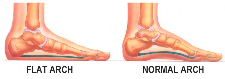
Surgical Treatment
For more chronic flatfoot pain, surgical intervention may be the best option. Barring other serious medical ailments, surgery is a good alternative for patients with a serious problem. There are two surgical options depending on a person?s physical condition, age and lifestyle. The first type of surgery involves repair of the PTT by transferring of a nearby tendon to help re-establish an arch and straighten out the foot. After this surgery, patients wear a non-weight bearing support boot for four to six weeks. The other surgery involves fusing of two or three bones in the hind foot below the ankle. While providing significant pain relief, this option does take away some hind foot side-to-side motion. Following surgery, patients are in a cast for three months. Surgery is an effective treatment to address adult-acquired flatfoot, but it can sometimes be avoided if foot issues are resolved early. That is why it is so important to seek help right away if you are feeling ankle pain. But perhaps the best way to keep from becoming flatfooted is to avoid the risk factors altogether. This means keeping your blood pressure, weight and diabetes in check.
What Are The Causes Of Heel Pain
Overview

Heel pain is a common foot condition. It's usually felt as an intense pain when using the affected heel. Heel Pain usually builds up gradually and gets worse over time. The pain is often severe and occurs when you place weight on the heel. In most cases, only one heel is affected, although estimates suggest that around a third of people have pain in both heels. The pain is usually worse first thing in the morning, or when you first take a step after a period of inactivity. Walking usually improves the pain, but it often gets worse again after walking or standing for a long time. Some people may limp or develop an abnormal walking style as they try to avoid placing weight on the affected heel.
Causes
There is no one cause of heel pain. Whole text books have been written on Disorders of the Heel. Some of the types of problems that can be seen in the heel include Heel spurs, these are small bony spurs that often develop on the bottom of the heel. They do not really cause any problems. It is only mentioned here as it is a common myth that they are a problem - almost always the pain associated with heel spurs is really plantar fasciitis. Plantar fasciitis is the most common cause of heel pain and is due to a strain of the long ligament along the bottom of the foot. The most symptom is pain when getting out of bed first thing in the morning ('post-static dyskinesia') A number of disease processes can uncommonly cause heel pain, such as rheumatoid arthritis, ankylosing spondylitis and gout. Stress fractures, which is an abnormal reaction of bone to stress can occur in those that are very active (eg athletes) or have weaker bones (eg osteoporosis) Pain at the back of the heel could be due to a number of problems, there could be a bursitis at the back of the heel bone (sometimes called 'Haglund's) there could be problems with the insertion of the achilles tendon, such as tendonitis or calcification. A 'stone' bruise is sometimes considered to be a cause of heel pain, its is simply a bruise of the bone. Another cause of heel pain is problems in the calf muscles that refer pain to the heel (myofascial trigger points) or pain referred from the lower back via the nerves from the back to the heel. Heel pain in children is usually due to severs disease or calcaneal apophysitis.
Symptoms
Initially, this pain may only be present when first standing up after sleeping or sitting. As you walk around, the muscle and tendon loosen and the pain goes away. As this problem progresses, the pain can be present with all standing and walking. You may notice a knot or bump on the back of the heel. Swelling may develop. In some cases, pressure from the back of the shoe causes pain.
Diagnosis
Your GP or podiatrist (a healthcare professional who specialises in foot care) may be able to diagnose the cause of your heel pain by asking about your symptoms and examining your heel and foot. You will usually only need further tests if you have additional symptoms that suggest the cause of your heel pain is not inflammation, such as numbness or a tingling sensation in your foot - this could be a sign of nerve damage in your feet and legs (peripheral neuropathy), your foot feels hot and you have a high temperature (fever) of 38C (100.4F) or above - these could be signs of a bone infection, you have stiffness and swelling in your heel - this could be a sign of arthritis. Possible further tests may include, blood tests, X-rays - where small doses of radiation are used to detect problems with your bones and tissues, a magnetic resonance imaging (MRI) scan or ultrasound scan, which are more detailed scans.
Non Surgical Treatment
Depending on the underlying cause, treatment can include. Rest from activities that stress the heel (such as running and jumping). Ice packs. Regular foot massage, concentrating on the arch of the foot. Professional strapping. A splint worn at night. Flexibility exercises. Ultrasound therapy. Anti-inflammatory medicine (topical or oral). Checking your posture and walking style, to correct imbalances and gait abnormalities that may contribute to the pain. Shoe inserts (orthoses) to help support the foot. In some cases, surgery may be recommended to treat conditions including neuroma, bursitis and heel spurs.
Surgical Treatment
Although most patients with plantar fasciitis respond to non-surgical treatment, a small percentage of patients may require surgery. If, after several months of non-surgical treatment, you continue to have heel pain, surgery will be considered. Your foot and ankle surgeon will discuss the surgical options with you and determine which approach would be most beneficial for you. No matter what kind of treatment you undergo for plantar fasciitis, the underlying causes that led to this condition may remain. Therefore, you will need to continue with preventive measures. Wearing supportive shoes, stretching, and using custom orthotic devices are the mainstay of long-term treatment for plantar fasciitis.
Prevention

The following steps will help prevent plantar fasciitis or help keep the condition from getting worse if you already have it. The primary treatment is rest. Cold packs application to the area for 20 minutes several times a day or after activities give some relief. Over-the-counter pain medications can help manage the pain, consult your healthcare professional. Shoes should be well cushioned, especially in the midsole area, and should have the appropriate arch support. Some will benefit from an orthotic shoe insert, such as a rubber heel pad for cushioning. Orthotics should be used in both shoes, even if only one foot hurts. Going barefoot or wearing slipper puts stress on your feet. Put on supportive shoes as soon as you get out of bed. Calf stretches and stretches using a towel (place the towel under the ball of your feet and pull gently the towel toward you and hold a few seconds) several times a day, especially when first getting up in the morning. Stretching the Achilles tendon at the back of the heel is especially important before sports, but it is helpful for nonathletes as well. Increasing your exercise levels gradually. Staying at a healthy weight. Surgery is very rarely required.
Heel pain is a common foot condition. It's usually felt as an intense pain when using the affected heel. Heel Pain usually builds up gradually and gets worse over time. The pain is often severe and occurs when you place weight on the heel. In most cases, only one heel is affected, although estimates suggest that around a third of people have pain in both heels. The pain is usually worse first thing in the morning, or when you first take a step after a period of inactivity. Walking usually improves the pain, but it often gets worse again after walking or standing for a long time. Some people may limp or develop an abnormal walking style as they try to avoid placing weight on the affected heel.
Causes
There is no one cause of heel pain. Whole text books have been written on Disorders of the Heel. Some of the types of problems that can be seen in the heel include Heel spurs, these are small bony spurs that often develop on the bottom of the heel. They do not really cause any problems. It is only mentioned here as it is a common myth that they are a problem - almost always the pain associated with heel spurs is really plantar fasciitis. Plantar fasciitis is the most common cause of heel pain and is due to a strain of the long ligament along the bottom of the foot. The most symptom is pain when getting out of bed first thing in the morning ('post-static dyskinesia') A number of disease processes can uncommonly cause heel pain, such as rheumatoid arthritis, ankylosing spondylitis and gout. Stress fractures, which is an abnormal reaction of bone to stress can occur in those that are very active (eg athletes) or have weaker bones (eg osteoporosis) Pain at the back of the heel could be due to a number of problems, there could be a bursitis at the back of the heel bone (sometimes called 'Haglund's) there could be problems with the insertion of the achilles tendon, such as tendonitis or calcification. A 'stone' bruise is sometimes considered to be a cause of heel pain, its is simply a bruise of the bone. Another cause of heel pain is problems in the calf muscles that refer pain to the heel (myofascial trigger points) or pain referred from the lower back via the nerves from the back to the heel. Heel pain in children is usually due to severs disease or calcaneal apophysitis.
Symptoms
Initially, this pain may only be present when first standing up after sleeping or sitting. As you walk around, the muscle and tendon loosen and the pain goes away. As this problem progresses, the pain can be present with all standing and walking. You may notice a knot or bump on the back of the heel. Swelling may develop. In some cases, pressure from the back of the shoe causes pain.
Diagnosis
Your GP or podiatrist (a healthcare professional who specialises in foot care) may be able to diagnose the cause of your heel pain by asking about your symptoms and examining your heel and foot. You will usually only need further tests if you have additional symptoms that suggest the cause of your heel pain is not inflammation, such as numbness or a tingling sensation in your foot - this could be a sign of nerve damage in your feet and legs (peripheral neuropathy), your foot feels hot and you have a high temperature (fever) of 38C (100.4F) or above - these could be signs of a bone infection, you have stiffness and swelling in your heel - this could be a sign of arthritis. Possible further tests may include, blood tests, X-rays - where small doses of radiation are used to detect problems with your bones and tissues, a magnetic resonance imaging (MRI) scan or ultrasound scan, which are more detailed scans.
Non Surgical Treatment
Depending on the underlying cause, treatment can include. Rest from activities that stress the heel (such as running and jumping). Ice packs. Regular foot massage, concentrating on the arch of the foot. Professional strapping. A splint worn at night. Flexibility exercises. Ultrasound therapy. Anti-inflammatory medicine (topical or oral). Checking your posture and walking style, to correct imbalances and gait abnormalities that may contribute to the pain. Shoe inserts (orthoses) to help support the foot. In some cases, surgery may be recommended to treat conditions including neuroma, bursitis and heel spurs.
Surgical Treatment
Although most patients with plantar fasciitis respond to non-surgical treatment, a small percentage of patients may require surgery. If, after several months of non-surgical treatment, you continue to have heel pain, surgery will be considered. Your foot and ankle surgeon will discuss the surgical options with you and determine which approach would be most beneficial for you. No matter what kind of treatment you undergo for plantar fasciitis, the underlying causes that led to this condition may remain. Therefore, you will need to continue with preventive measures. Wearing supportive shoes, stretching, and using custom orthotic devices are the mainstay of long-term treatment for plantar fasciitis.
Prevention
The following steps will help prevent plantar fasciitis or help keep the condition from getting worse if you already have it. The primary treatment is rest. Cold packs application to the area for 20 minutes several times a day or after activities give some relief. Over-the-counter pain medications can help manage the pain, consult your healthcare professional. Shoes should be well cushioned, especially in the midsole area, and should have the appropriate arch support. Some will benefit from an orthotic shoe insert, such as a rubber heel pad for cushioning. Orthotics should be used in both shoes, even if only one foot hurts. Going barefoot or wearing slipper puts stress on your feet. Put on supportive shoes as soon as you get out of bed. Calf stretches and stretches using a towel (place the towel under the ball of your feet and pull gently the towel toward you and hold a few seconds) several times a day, especially when first getting up in the morning. Stretching the Achilles tendon at the back of the heel is especially important before sports, but it is helpful for nonathletes as well. Increasing your exercise levels gradually. Staying at a healthy weight. Surgery is very rarely required.